
Jul 30
Jul 30
Jul 29
Jul 22
Jul 19
Jul 17
Late-night sugar consumption may seem harmless, but biologically it creates one of the highest-risk environments for enamel erosion and bacterial activity. Saliva flow decreases during sleep, weakening the mouth’s natural defense against acid production and plaque formation. Harmful oral bacteria metabolize sugar and release acids that continue damaging tooth surfaces for hours while the body rests. Over time, repeated late-night sugar intake contributes to cavities, gum inflammation, and systemic inflammatory responses linked to broader health conditions. Understanding the science behind night-time oral ecology helps individuals adopt protective habits that safeguard both dental integrity and overall wellness.
Oral physiology changes significantly during sleep. Salivary flow — the mouth’s primary protective mechanism — drops dramatically. Saliva normally:
• Neutralizes bacterial acids
• Flushes away food particles
• Supplies minerals that repair enamel
When sugar is consumed shortly before sleep:
• Acid neutralization is reduced
• Sugar residue remains longer on tooth surfaces
• Plaque biofilm becomes more active
This combination allows enamel damage to continue uninterrupted for several hours. Repeated exposure compounds risk even when overall sugar intake appears moderate.
The oral microbiome contains species that metabolize carbohydrates into acids, especially Streptococcus mutans, a major contributor to tooth decay.
During night conditions, these bacteria:
• Multiply more rapidly
• Produce sustained acid output
• Penetrate enamel mineral layers
• Initiate demineralization cycles
Because brushing often does not occur afterward, the bacterial metabolic window extends throughout sleep. This is why dental professionals consistently recommend avoiding food or beverages (except water) before bed.
Oral health is closely linked with overall physiology. Chronic late-night sugar exposure can contribute to:
• Gum inflammation associated with cardiovascular stress markers
• Increased systemic inflammatory signaling
• Blood glucose fluctuation that may impact sleep regulation
• Microbial imbalance affecting immune response
While occasional indulgence is unlikely to cause harm, repeated behavioral patterns influence long-term metabolic and inflammatory outcomes.
Behavioral adjustment is the most effective prevention method.
Evidence-based actions include:
• Brushing thoroughly before sleep
• Rinsing with water if brushing isn’t possible
• Choosing low-sugar, tooth-neutral snacks
• Avoiding prolonged sipping or grazing
Tooth-friendly options:
• Cheese
• Nuts
• Plain yogurt
These foods stimulate saliva or provide protective minerals.
Fatigue often reduces brushing quality during evening routines.
Coverage gaps commonly occur along:
• Gumlines
• Inner tooth surfaces
• Posterior molars
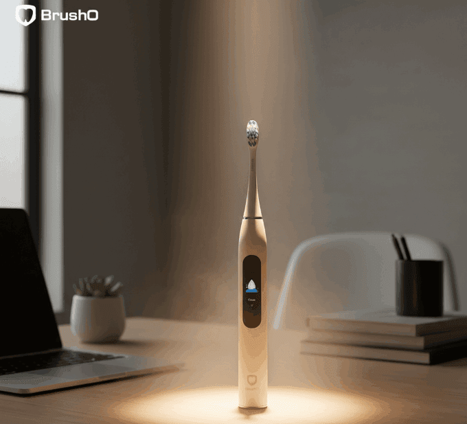
Smart brushing platforms such as BrushO support consistent technique by:
• Monitoring brushing coverage
• Reinforcing routine consistency
• Guiding full-mouth cleaning
This helps reduce bacterial survival windows before sleep begins.
Midnight sugar consumption shifts the oral environment in favor of acid-producing bacteria at a time when natural defenses are weakest. Over time, this habit contributes to enamel degradation, gum irritation, and broader health implications linked to inflammation and metabolic balance. Preventive brushing, informed dietary choices, and consistent hygiene monitoring remain the most effective strategies for maintaining oral stability despite occasional cravings.
Feb 9
Feb 23
Jul 30
Jul 30
Jul 29
Jul 22
Jul 19
Jul 17

Hyperdontia is the presence of one or more teeth in excess of the normal dental formula, and the resulting teeth are described as supernumerary. The condition is common enough to appear in routine practice but variable enough in presentation that the decision to intervene is rarely straightforwar...

Few patients complete orthodontic treatment without asking whether their teeth will move back. The honest answer is that they will, to some degree, in the majority of cases. Relapse is not a failure of treatment but a predictable biological response, and understanding why it happens is the basis ...

The mandible is the largest and most frequently fractured bone of the facial skeleton, and its management sits at the interface between trauma surgery and restorative dentistry. Treatment has shifted over four decades from prolonged maxillomandibular fixation towards rigid internal fixation with ...

Chronic liver disease affects hundreds of millions of people worldwide, and its prevalence continues to rise with the epidemic of metabolic liver disease. The liver is central to haemostasis, drug metabolism and immune competence, and its failure therefore alters the risk profile of almost every ...

Excessive gingival display, commonly described as a gummy smile, is one of the most frequent aesthetic concerns presented to dental practitioners. The condition is not a single entity but a clinical presentation that can arise from skeletal, gingival, muscular or dental factors, and the treatment...

Drug-induced gingival overgrowth is a well-recognised adverse effect of several widely prescribed medications, and it remains one of the most common drug-related findings in periodontal practice. The condition produces firm, fibrous enlargement of the gingiva that can interfere with speech, masti...

Denture stomatitis is an inflammatory condition of the denture-bearing mucosa that affects a large proportion of removable prosthesis wearers, yet it is frequently dismissed as a cosmetic annoyance. The condition is driven by a Candida biofilm on the fitting surface of the prosthesis, and it is m...

Chronic kidney disease affects an estimated 850 million people worldwide, and the number of patients surviving on dialysis or with a renal transplant continues to rise. These patients carry an oral disease burden that is substantially higher than that of the general population, and their medical ...

Angular cheilitis is an inflammatory lesion of the labial commissures that presents with erythema, fissuring and crusting at the corners of the mouth. It is one of the most common conditions encountered in general dental practice, yet it is frequently treated as a simple fungal infection and allo...

Adverse reactions to dental materials are uncommon but they are not rare, and they are frequently misdiagnosed because the oral mucosa responds to allergens in a limited number of ways. A burning sensation, a persistent ulcer or an unexplained area of erythema may all represent a hypersensitivity...

BrushO AI Electric Toothbrush offers real-time brushing guidance, pressure feedback, coverage tracking, and app-based progress reports for a cleaner daily routine.
BrushO AI Electric Toothbrush offers real-time brushing guidance, pressure feedback, coverage tracking, and app-based progress reports for a cleaner daily routine.


Address: RM 1427, 14/F RADIO CITY 505, HENNESSY RD CAUSEWAY BAY, HONG KONG
Code: 000000
Phone: +852-53758652
Email: customerservice@brusho.com