
Jul 30
Jul 30
Jul 29
Jul 22
Jul 19
Jul 17
Tooth loss is often viewed as a localized dental issue, but its effects extend far beyond the mouth. Each tooth plays a vital role in maintaining jawbone density, facial symmetry, and structural support for soft tissue. When teeth are lost, the surrounding bone gradually resorbs due to reduced stimulation, altering facial contours and accelerating visible aging. Understanding the biological mechanisms behind bone remodeling after tooth loss highlights the importance of preventive oral care, early intervention, and advanced monitoring technologies that support long-term structural health.
Teeth are not just functional tools for chewing — they are biomechanical stimulators of the jawbone. Every bite and chewing motion transfers force through the tooth root into the surrounding bone tissue. This mechanical stimulation signals the body to maintain bone density through continuous remodeling.
Healthy teeth help preserve:
• Jawbone volume
• Facial symmetry
• Muscle attachment stability
• Soft tissue support
Without this stimulation, bone maintenance mechanisms slow down.
When a tooth is lost, the alveolar bone that once supported it no longer receives mechanical stimulation. The body interprets this as unnecessary tissue and gradually reabsorbs it. This process can start within months and continue for years.
• Reduced bone thickness
• Decreased bone height
• Altered jaw structure
Bone loss is permanent without medical intervention.
As bone volume decreases, external facial changes may appear:
• Sunken cheeks
• Thinner lips
• Shortened lower face height
• Wrinkling around the mouth
• Reduced chin projection
These changes contribute to premature facial aging and altered expression dynamics.
Tooth loss affects neighboring teeth and bite mechanics:
• Adjacent teeth may shift
• Opposing teeth may over-erupt
• Chewing force distribution becomes uneven
This further accelerates structural imbalance and joint strain.
Beyond aesthetics, bone changes influence:
• Speech articulation
• Chewing efficiency
• Digestive effectiveness
• Temporomandibular joint (TMJ) stability
Maintaining dental integrity is therefore a whole-system health priority.
Periodontal disease is a major contributor to tooth loss. Controlling bacterial buildup helps preserve supporting structures.
Aggressive brushing or missed zones increases gum and bone risk.
Early detection of inflammation prevents progression to irreversible structural damage.
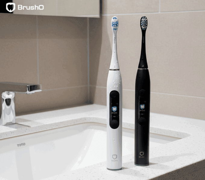
AI-guided toothbrush technologies like BrushO assist users in maintaining protective habits that reduce tooth loss risk.
Prevents gum trauma that contributes to recession and instability.
Ensures complete plaque removal across all surfaces.
Identifies behavioral patterns linked to oral health decline.
Adapt intensity to protect sensitive or at-risk areas.
These tools support long-term preservation of dental and bone structure through consistent daily feedback.
Dental professionals may recommend:
• Implants
• Bridges
• Dentures
These interventions restore function and partially preserve bone stimulation, though prevention remains the optimal strategy.
Tooth loss triggers structural changes that extend far beyond missing teeth. Reduced bone stimulation leads to jaw resorption, facial contour alteration, and functional complications. Recognizing this connection reinforces the importance of preventive oral hygiene and consistent monitoring. Combining daily care practices with intelligent brushing guidance — such as provided by BrushO — helps reduce the risk of periodontal decline and supports lifelong structural health. Oral health preservation is not only about maintaining a smile — it is about protecting the architecture of the face itself.
Jul 30
Jul 30
Jul 29
Jul 22
Jul 19
Jul 17

Hyperdontia is the presence of one or more teeth in excess of the normal dental formula, and the resulting teeth are described as supernumerary. The condition is common enough to appear in routine practice but variable enough in presentation that the decision to intervene is rarely straightforwar...

Few patients complete orthodontic treatment without asking whether their teeth will move back. The honest answer is that they will, to some degree, in the majority of cases. Relapse is not a failure of treatment but a predictable biological response, and understanding why it happens is the basis ...

The mandible is the largest and most frequently fractured bone of the facial skeleton, and its management sits at the interface between trauma surgery and restorative dentistry. Treatment has shifted over four decades from prolonged maxillomandibular fixation towards rigid internal fixation with ...

Chronic liver disease affects hundreds of millions of people worldwide, and its prevalence continues to rise with the epidemic of metabolic liver disease. The liver is central to haemostasis, drug metabolism and immune competence, and its failure therefore alters the risk profile of almost every ...

Excessive gingival display, commonly described as a gummy smile, is one of the most frequent aesthetic concerns presented to dental practitioners. The condition is not a single entity but a clinical presentation that can arise from skeletal, gingival, muscular or dental factors, and the treatment...

Drug-induced gingival overgrowth is a well-recognised adverse effect of several widely prescribed medications, and it remains one of the most common drug-related findings in periodontal practice. The condition produces firm, fibrous enlargement of the gingiva that can interfere with speech, masti...

Denture stomatitis is an inflammatory condition of the denture-bearing mucosa that affects a large proportion of removable prosthesis wearers, yet it is frequently dismissed as a cosmetic annoyance. The condition is driven by a Candida biofilm on the fitting surface of the prosthesis, and it is m...

Chronic kidney disease affects an estimated 850 million people worldwide, and the number of patients surviving on dialysis or with a renal transplant continues to rise. These patients carry an oral disease burden that is substantially higher than that of the general population, and their medical ...

Angular cheilitis is an inflammatory lesion of the labial commissures that presents with erythema, fissuring and crusting at the corners of the mouth. It is one of the most common conditions encountered in general dental practice, yet it is frequently treated as a simple fungal infection and allo...

Adverse reactions to dental materials are uncommon but they are not rare, and they are frequently misdiagnosed because the oral mucosa responds to allergens in a limited number of ways. A burning sensation, a persistent ulcer or an unexplained area of erythema may all represent a hypersensitivity...

La brosse à dents électrique BrushO avec IA offre un guidage en temps réel, un retour sur la pression, un suivi de la couverture et des rapports de progression dans l’app pour une routine quotidienne plus propre.
La brosse à dents électrique BrushO avec IA offre un guidage en temps réel, un retour sur la pression, un suivi de la couverture et des rapports de progression dans l’app pour une routine quotidienne plus propre.


Address: RM 1427, 14/F RADIO CITY 505, HENNESSY RD CAUSEWAY BAY, HONG KONG
Code: 000000
Phone: +852-53758652
Email: customerservice@brusho.com