
Jul 30
Jul 30
Jul 29
Jul 22
Jul 19
Jul 17
Many people become concerned when they notice raised bumps or nodules at the back of their tongue. In most cases, these structures are normal anatomical features known as papillae that support taste perception, digestion, and immune defense. However, changes in appearance, sensation, or persistence can signal irritation, infection, or nutritional imbalance. Understanding the biological role of tongue structures and maintaining effective hygiene practices helps prevent unnecessary anxiety while promoting oral wellness. This article explains tongue anatomy, warning indicators, and evidence-based care strategies that support both oral and systemic health.
The surface of the tongue is not smooth by design. It contains specialized structures called papillae, which perform critical biological functions.
• Hosting taste receptors
• Assisting with food manipulation and swallowing
• Supporting oral immune surveillance
• Helping maintain microbial balance
Circumvallate Papillae
Large dome-shaped bumps arranged in a V-shape at the back of the tongue (typically 8–12). These are the bumps most people notice and are usually normal.
Fungiform Papillae
Smaller mushroom-shaped structures near the tip and sides, containing taste buds.
Foliate Papillae
Located along the rear sides; often more visible in younger individuals.
Filiform Papillae
Most abundant type; provides texture and grip for food but does not contain taste buds.
Seeing bumps at the back of the tongue is therefore typically a sign of healthy anatomy rather than disease.
Although papillae are normal, certain changes should be monitored.
• Mechanical irritation from hot or spicy foods
• Minor trauma from biting
• Temporary inflammation
• Persistent redness or swelling
• White or yellow coating
• Painful nodules
• Bleeding or ulceration
• Changes lasting more than two weeks
These symptoms may reflect:
• Oral infections
• Nutritional deficiencies
• Microbial imbalance
• Chronic irritation
Professional evaluation is recommended when abnormalities persist.
The tongue hosts a significant portion of oral bacteria and contributes to the overall oral microbiome.
• Reduces odor-causing bacteria
• Supports microbial balance
• Improves taste perception
• Lowers plaque transfer to teeth
• Gently brush or scrape the tongue daily
• Rinse after meals
• Maintain hydration
• Use antimicrobial rinses when appropriate
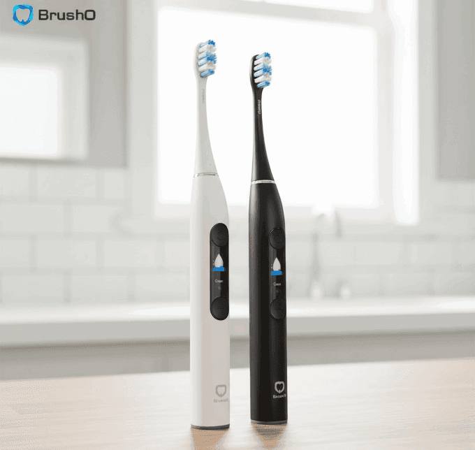
Advanced oral care tools such as BrushO assist in maintaining comprehensive cleaning by guiding full-mouth coverage beyond just tooth surfaces.
Oral tissues often reflect broader physiological conditions.
• White coating — immune stress or microbial imbalance
• Smooth red tongue — vitamin deficiency signals
• Swollen papillae — inflammatory response
• Persistent lesions — require clinical assessment
The tongue functions as an early biological indicator, highlighting the importance of consistent monitoring.
Bumps at the back of the tongue are typically normal papillae essential for taste, digestion, and immune defense. Awareness of anatomical variation prevents unnecessary concern, while understanding warning signs supports early intervention when needed. Integrating proper tongue hygiene into daily oral care routines contributes to both oral microbiome balance and overall wellness. Smart oral care technologies like BrushO help ensure comprehensive cleaning coverage, supporting healthier habits and improved long-term oral outcomes.
Feb 20
Feb 19
Jul 30
Jul 30
Jul 29
Jul 22
Jul 19
Jul 17

A lateral cephalometric radiograph is only as useful as the analysis applied to it. The same tracing can be read as a skeletal discrepancy by one method and as a dental compensation by another, and the conclusions reached about extraction, surgery or growth modification may differ accordingly. Un...

Premature fusion of the cranial sutures produces a group of conditions in which the skull, the midface and the occlusion develop abnormally, and the dental team is central to long-term care. Apert and Crouzon syndromes are the best known craniosynostosis syndromes with major oral consequences. Bo...

A dense opacity on a panoramic radiograph above the molar roots or within the nasal cavity is a familiar finding that is often dismissed as an incidental artifact. When that opacity proves to be an antrolith or a rhinolith, the discovery explains years of unilateral obstruction, discharge and fac...

A firm, board-like swelling at the angle of the jaw that slowly breaks down into multiple draining sinuses should raise suspicion of cervicofacial actinomycosis, a chronic bacterial infection that is easily mistaken for a tumor. Nocardiosis, caused by related filamentous organisms, can produce a ...

Enlargement of the jaw, widening spaces between the teeth and a slowly changing facial profile may bring a patient to the dental office years before the underlying endocrine disease is diagnosed. Acromegaly and Cushing syndrome both leave recognizable signatures in the craniofacial region, and th...

A slowly growing, painless lump at the angle of the jaw in a middle-aged or older man who smokes is the classic presentation of a Warthin tumor. Formally called papillary cystadenoma lymphomatosum, this benign neoplasm is one of the most common tumors of the parotid gland and the second most comm...

Painful ulcerated gums that bleed easily, a foul metallic taste and severe mouth odor can signal Vincent stomatitis, an acute fusospirochetal necrotizing infection of the gingival and oral tissues. First described during wartime and historically called trench mouth, the disease still occurs today...

A painful swelling under the jaw that appears at mealtimes and subsides slowly afterward is the classic symptom of a salivary stone blocking a duct. Sialolithiasis, the formation of calcified stones in the salivary glands, and sialodochitis, the inflammation of the ducts that follows, are common ...

Paget disease of bone, also called osteitis deformans, is a chronic disorder of bone remodeling that can silently affect the skull and the jaws. In the maxillofacial region it produces progressive bony enlargement, distinctive cotton-wool radiographs and dental complications such as hypercementos...

The sudden appearance of widespread, painful oral erosions with crusted lips can be alarming, and it may represent either erythema multiforme or a drug-induced severe reaction such as Stevens-Johnson syndrome. Both belong to a spectrum of immune-mediated mucocutaneous disorders, and both can begi...

BrushO AI इलेक्ट्रिक टूथब्रश रियल-टाइम ब्रशिंग गाइड, प्रेशर फीडबैक, कवरेज ट्रैकिंग और ऐप-आधारित प्रगति रिपोर्ट देता है, ताकि आपकी दैनिक दिनचर्या अधिक साफ और समझने योग्य बने।
BrushO AI इलेक्ट्रिक टूथब्रश रियल-टाइम ब्रशिंग गाइड, प्रेशर फीडबैक, कवरेज ट्रैकिंग और ऐप-आधारित प्रगति रिपोर्ट देता है, ताकि आपकी दैनिक दिनचर्या अधिक साफ और समझने योग्य बने।


Address: RM 1427, 14/F RADIO CITY 505, HENNESSY RD CAUSEWAY BAY, HONG KONG
Code: 000000
Phone: +852-53758652
Email: customerservice@brusho.com